
Swapping glass slides for DNA-encoded viruses: Advanced tools to study sugar interactions in health and disease
GlycoNet researchers at the University of Alberta have developed two cutting-edge tools to help fine-tune our
understanding of sugar molecules called glycans.
By Aalyssa Atley • Posted: 16 December, 2024
Glycans—complex sugar molecules that decorate the surfaces of cells—play a pivotal role in everything from immune response to disease progression, yet their interactions remain some of the most challenging mysteries in biology.
Upgrading glass slides to DNA-encoded viruses
For the past few decades, glycomics—the study of glycans or sugars in biological processes—has relied heavily on analysis using traditional microarrays (similar to microscope slides). By attaching diverse glycans to glass slides, researchers can observe which molecules bind to specific glycans.
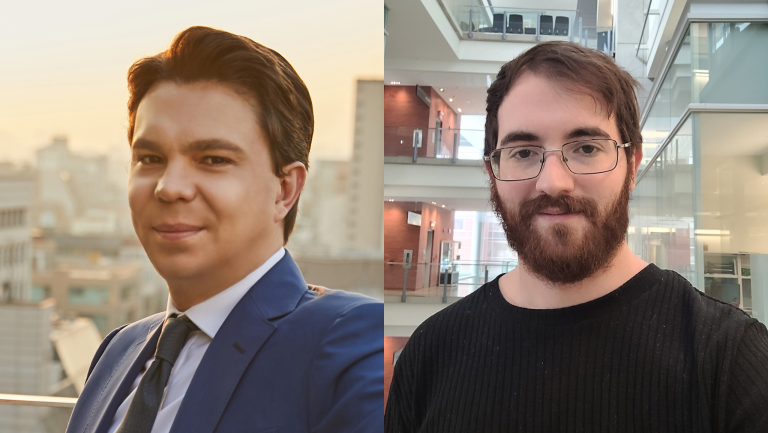
“However, if you want to study glycan interactions inside living organisms, glass slides fall short,” explains Ratmir Derda, a GlycoNet investigator and professor at the University of Alberta. “Glass slides can’t replicate the dynamic environment of a living organism.”
Derda saw an opportunity to address this gap using harmless viruses called bacteriophages, or phages. As an alternative to printing molecules on glass slides, phages can be engineered to display specific molecules on their surface.
“Bacteriophages are basically like a glass slide,” Derda notes, “but they’re so small and non-invasive that you can actually inject them into the body.”
Each phage also carries a unique DNA ‘barcode’ that enables scientists to track and identify them, making them a useful tool for studying biological environments.
With contributions from GlycoNet investigators Matthew Macauley et à Lara Mahal at the University of Alberta, Derda’s team adapted this versatile phage technology to create two complementary platforms: the Liquid Glycan Array (LiGA), and most recently, the Liquid Lectin Array (LiLA).
“Combining our expertise in the family of Siglec receptors—sialic acid-binding lectins—with the Derda lab’s unique expertise in genetic encoding has allowed us to develop tools that advance the boundaries of the glycosciences,” says Macauley.
Two sides of the same coin
LiGA and LiLA are built on the same underlying technology but with different applications. LiGA displays glycans on phages, enabling the detection of glycan-binding receptors. In contrast, LiLA displays lectins, allowing researchers to explore glycan profiles on cells.
“They’re like two sides of the same coin,” says Derda. “Think of glycans and lectins as keys and locks. LiGA helps identify which keys (glycans) fit which locks (lectins), while LiLA does the reverse.”
Although LiGA and LiLA share similarities, the creation of LiLA was more challenging because lectins are larger than glycans and more difficult to attach to phages.
“We had to devise a system different from the chemistry used to attach glycans to phages,” says Guilherme Lima, a postdoctoral researcher in the Derda lab, who played a lead role in solving this challenge.
“By adapting some existing technology, we created a kind of ‘glue’ to attach the proteins to the phage.”
Magnifying subtle changes
As complementary tools, LiGA and LiLA provide a more agile approach to studying how glycans and proteins interact within living systems. They also have the capacity to amplify subtle details that enhance our understanding of biology.
One key advantage of LiGA and LiLA is their ability to precisely control how glycans and lectins are displayed or spatially presented to each other, revealing new dimensions of how these interactions influence biology and health.
For example, in their recent paper on LiLA, the research team found that adjusting the density of lectins on phages allowed them to distinguish between high and low levels of a sugar called sialic acid, which is often found in higher amounts on cancer cells.
“These changes are very subtle, but our platform is able to detect and amplify those changes, so we can understand better,” explains Lima.
This capability enabled the research team to identify a specific configuration of lectins that binds only to cancer cells with high sialic acid density while ignoring the lower density of sialic acid on normal tissue—a finding that could lead to more targeted therapies.
“The same principle also applies to LiGA for density,” adds Lima. “The number of glycans present on the phages is important.”
Opening doors in biology and health
“If we want to direct therapies to cells and organs, and the major component of the cell surface is a sugar, but we tend to ignore it and just focus on the minor components, then we are kind of robbing ourselves of many opportunities,” says Derda.
Traditional tools used in other areas of biology have struggled with the complexity of glycans. LiGA and LiLA are a step forward to helping us understand the roles of glycans in greater depth.
For instance, LiLA was also able to detect that cancer stem-like cells exhibit different glycosylation profiles than other non-stem cancer cells, an observation that could help improve the design of future cancer treatments.
“If you can kill those cancer stem cells—the ones that actually make more of the cancer cells—then there could be less relapse after treatment,” says Lima.
The applications of LiGA and LiLA extend beyond cancer, as glycan-lectin interactions play critical roles in numerous biological processes and diseases, from immune response to infection pathways. Understanding the nuances of these interactions opens up opportunities for developing new diagnostics and treatments.
Access to LiGA and LiLA
As the research team continues expanding the applications of these platforms and refining their use in living systems, Derda also plans to start a new company to make LiGA and LiLA more widely available.
LiGA is already available for use as part of the offerings of Les Services intégrés de GlycoNet, which provides academic and industry clients with access to specialized glycomics tools, expertise and services.
In addition to their newest paper on LiLA, the research team recently published a detailed protocol on using LiGA.
Funding for the development of LiGA and LiLA was provided by Alberta Innovates, the Canadian Institutes of Health Research, GlycoNet, and the Natural Sciences and Engineering Research Council of Canada. Critical glycan components were also sourced from the Consortium of Functional Glycomics and several other researchers.